기사본문
Tomocube’s revolutionary holotomography microscope opens up a new chapter for real-time live cell imaging
입력 2017-09-19 10:31 수정 2017-09-19 10:31
바이오스펙테이터 Joungmin Cho 기자

"Let us imagine that a researcher intends to observe target living cells using traditional microscopy techniques. The researcher needs to perform the following cumbersome and time-consuming procedures: (1) slide preparation (specimen fixation and tissue processing; 30 minutes to 1 hour), (2) stages evolved for the specific reaction via antibody incubation at the target site (8 to 12 hours), and (3) macroscopic observation of antibody retrieval following the secondary antibody treatment with fluorescent proteins (2 to 4 hours). Even in the mid-process, the washing process is indispensable for preparation of the appropriate solutions (3 hours). After 48 hours, the researcher can analyze desired cells, but the cells are inevitably dead or significantly affected due to the use of labeling agents.”
After the development of the microscope in the 17th century, microscopic analysis of biological cells and tissues has become available as indispensable tools in biomedical research, but a real-time exploration of living cells remains a daunting task. Although there is a technique of inserting fluorescence-expressing genes into living cells, it also requires genetic modification, preventing from the study of intact cells. It becomes big limitations, especially when cells should be treated in their intact conditions, such as stem cell research or immunotherapy.
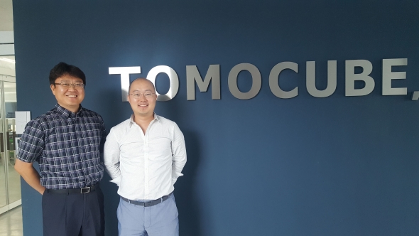
▲(left)Ki-hyun Hong, CEO of Tomocube (right) Young-keun Park, CTO
To solve the problem, Tomocube introduced a state-of-art 3D holographic microscope that allows real-time label-free visualization of biological cells and tissues without any sample preparation or labeling.
Professor Yong-Keun Park of the Physics Department at the Korea Advanced Institute of Science and Technology (KAIST), a co-founder and Chief Technical Officer of Tomocube, has been leading this field. Prof. Park colleagues developed a holotomography (HT)-1 technology which is designed to observe living cells by employing the principle of computed tomography (CT).
Professor Park commented that “Our new HT-1 microscope enables real-time measurements of unlabeled live cells and accurate quantitative imaging analysis. Thus, we expect that this tomographic microscopy will be the best suitable model in the age of artificial intelligence (AI).” In the series of recent publications, hHis team e confirmed has demonstrated that HT-1 has potentials for the study of live cells in real time for various applications, including hematology, infectious diseases, cell biology, neuroscience, and cytotoxicity.
“Scientists have further expanded the potential of fluorescent proteins, from basic biology to molecular diagnosis,” Ki-hyun Hong, CEO of Tomocube, explained. “Our HT-1 technology will set a new paradigm for medical diagnosis.”.
Researchers want real-time dynamics of living cells. It is also necessary to process the image data quantitatively in order to treat the observation results as meaningful data.
Professor Park stated that “To meet their needs, such as confocal microscopy and electron microscopy. However, none of the previous approaches provides imaging of unlabeled cells, particularly in 3D and a quantitative manner.”
The imaging contrast of holotomography comes from refractive index (RI), an intrinsic optical parameter of a material. In holotomography, the 3D image of a living cell can be obtained by measuring the 3D RI distribution of a sample. Professor Park noted that “When light passes through an object, diffraction occurs according to the object’s RI distribution. The diffraction patterns of a sample are holographically measured with various illumination patterns, from which the 3D RI tomogram of the sample is reconstructed. The reconstructed 3D RI map provides both structural and chemical information regarding the cell, because the RI information of a cell can be translated into protein concentration.”.
Tomocube’s technology enables real-time viewing and precise measurement of living cells without using any labeling agents. Morphologies of cell membrane as well as subcellular organelles can be seen. Furthermore, the mass of these subcellular structures can also be calculated.
Following the first commercialization of HT-1, Tomocube introduced ‘HT-2’, another holotomography series equipped with the 3D fluorescence imaging technology; thus, researchers have easier access to HT-2.
Professor Park stated that “HT-2 is an upgraded version designed to provide existing fluorescent imaging to allow researchers perform correlative studies using both the fluorescence and holography images of cells, which provide complementary information."
Tomocube announced its long-term plan to embrace ‘Big Data,’, as part of efforts to construct a large library that could manage all data of biological cells obtained by researchers who use the HT microscope, and to eventually utilize these data for more rapid and precise diagnosis of diseases..
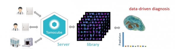
Professor Park added that “The strong point of Tomocube’s technology toward diagnosis based on AI is the use of intrinsic RI information. Cell images can be obtained without procedures and can be quantitatively measured in a highly reproducible manner, which suggests the on-site and precise diagnosis.”
Recently, medical diagnosis based on CT or MRI images have been empowered with AI; several expertsit is anticipated that large portions of clinicians’ own experience and interpretation can be eventually replaced by algorithms and big data. However, in histology or cytology, one of the challenges towardof AI-based medical diagnosis is how to acquire a largesignificant amount of gooduseful data. In particular, current labeling methods in imaging tissues and cells are time-consuming, and more importantly, now provide quantitative imaging results. Sometimes fluorescence or dye labeling image results are not producible, or cause damages to samples.
In contrast, Tomocube’s technology may enables rapid on-site diagnosis, because it measured quantitative RI tomograms of cells and tissues. Professor Park stated that “We can expect the best synergistic effects, when our technology is combined with AI.”.
CEO Hong stated that “A number of domestic and international hospitals and research centers, such as Seoul National University, Asan Medical Center, MIT/Harvard medical school, Texas Medical Institute, and German cancer research center, are using our technology for various research topics, and it will provide us numerous cell and tissue image data in turn”. In addition, Tomocube is currently expanding its sales network covering major countries.
Tomocube aims to provide a novel better medical diagnosis of human diseases criteria by bringing innovations in seeing live cells using holography and AI.